
Researchers use a temporary “liver factory” to restore T cell strength in older mice (Figure 1).

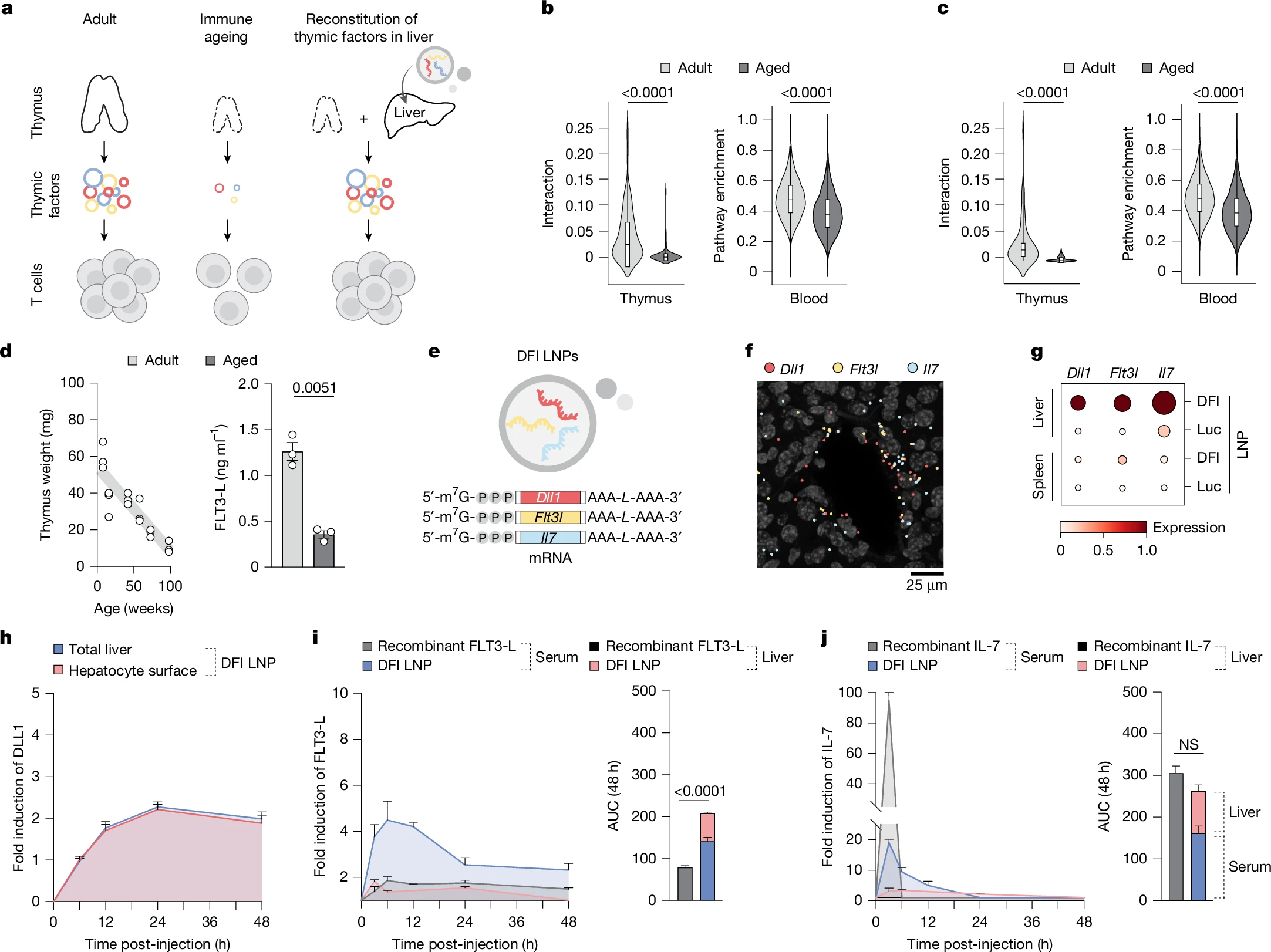
Figure 1: Hepatic reconstitution of declining T cell signalling factors to restore immune signalling in ageing. a, Overview of the approach to restore age-declining immune trophic cues by hepatic expression of Dll1, Flt3l and Il7 mRNAs. b, Spatial ligand–receptor interactions between thymic cortical epithelial cells (cTECs) and thymocytes decline with age (left), and ssGSEA shows reduced Notch pathway activity in circulating T cells (right). n = 47 spatial arrays and 96,683 blood T cell transcriptomes across 21 ages. Data are represented as violin plots with median + interquartile range. Statistical significance was determined by Mann–Whitney tests. c, cTEC–T cell IL-7 interaction (left) and downstream pathway in circulating T cell (right) activities are likewise diminished with age. n = 47 spatial arrays and 96,683 blood T cell transcriptomes across 21 ages. Data are represented as violin plots with median + interquartile range. Statistical significance was determined by Mann–Whitney tests. d, Thymus weight decreases with age (n = 18; 3 per timepoint). Interstitial FLT3-L levels are reduced in aged thymus by ELISA (n = 3 per group). Data are mean ± s.e.m.; statistical significance was determined by a two-tailed unpaired Student’s t-test. e, mRNA (DFI; Dll1, Flt3l and Il7) constructs formulated in SM-102 LNPs. f, Representative RIBOmap images 6 h post-DFI show robust ribosome-bound transcripts in the liver. A representative image from three imaged DFI-treated animals is shown. g, Single-cell quantification: translating Dll1, Flt3l and Il7 in the liver and spleen by RIBOmap (n = 1 for Luc and n = 3 for DFI). h, Immunofluorescence of DLL1 protein over 0–48 h after 5 µg DFI reveals transient induction in total liver and hepatocyte surface (phalloidin co-stain). Fold induction from baseline (0 h) is shown. n = 32 fields of view from n = 3 animals per time point per condition. Data are mean ± s.e.m. i, ELISA for FLT3-L levels in serum and the liver after 10 µg recombinant FLT3-L or 5 µg DFI at 3–48 h. Liver concentrations were normalized to liver weight; fold change from 0 h is shown. n = 3 animals per time point per compartment per condition. Data are mean ± s.e.m.; area under the cover (AUC) over 48 h compared by a two-tailed unpaired Student’s t-test. j, ELISA for IL-7 in serum and the liver after 10 µg recombinant IL-7 or 5 µg DFI at 3–48 h with liver normalization and fold change from 0 h. n = 3 animals per time point per compartment per condition. Data are mean ± s.e.m.; 48-h AUC compared by a two-tailed unpaired Student’s t-test. NS, not significant.
- DLL1, involved in T cell lineage commitment
- FLT-3, which supports immune progenitor survival
- IL-7, a key cytokine for T cell maintenance
- Larger and more diverse T cell populations
- Improved T cell function