
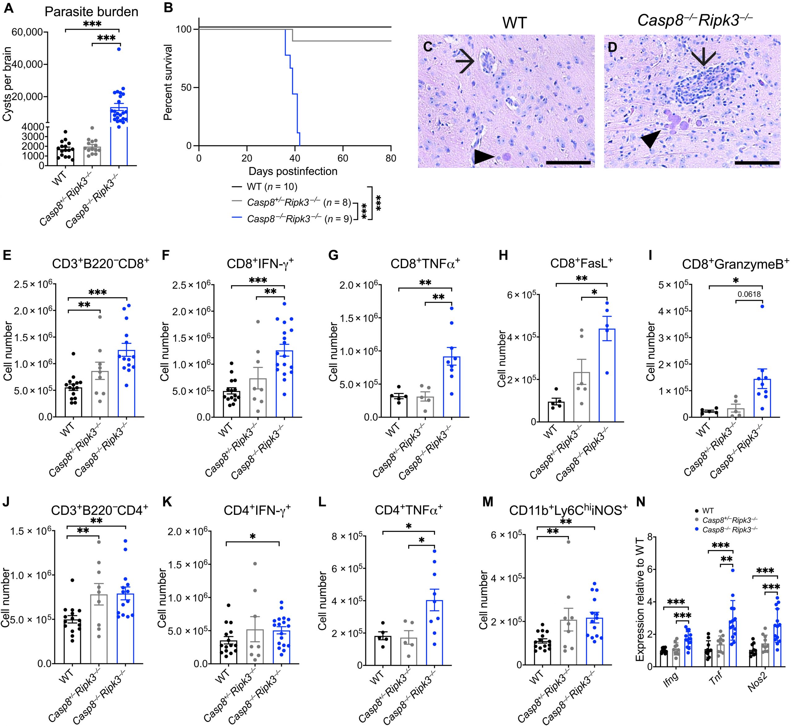
New research reveals that CD8⁺ T cells rely on caspase-8 to control Toxoplasma infection in the brain (Figure 1).

Figure 1: Caspase-8 is essential for controlling T. gondii infection in the brain, but Casp8 deficiency did not affect the TH1 cell immune response. Casp8−/−Ripk3−/−, Casp8+/−Ripk3−/−, and WT C57BL/6 mice were infected with 10 cysts of the Me49 strain of T. gondii intraperitoneally and analyzed at 4 wpi. (A) Cyst burden was quantified by light microscopy. (B) Survival curve of infected WT, Casp8+/−Ripk3−/−, and Casp8−/−Ripk3−/− mice. Representative H&E image of (C) WT and (D) Casp8−/−Ripk3−/− brain at 4 wpi. Arrows indicate inflamed blood vessels, and arrowheads indicate T. gondii cysts. Scale bars, 50 μm. Brain immune cell populations were quantified by spectral flow cytometry at 4 wpi: (E) CD8+ T cells, (F) CD8+IFN-γ+ T cells, (G) CD8+TNFα+ T cells, (H) CD8+FasL+ T cells, (I) CD8+GranzymeB+ T cells, (J) CD4+ T cells, (K) CD4+IFN-γ+ T cells, (L) CD4+TNFα+ T cells, and (M) infiltrating iNOS+ monocytes (CD45hiCD11b+Ly6G−Ly6ChiNOS+). (N) Gene expression was measured by RT-qPCR for Ifng, Tnf, and Nos2. Statistical significance determined by randomized block analysis of variance (ANOVA) and least-squares means: (A) WT C57BL/6 (n = 16), Casp8+/−Ripk3−/− (n = 14), and Casp8−/−Ripk3−/− (n = 22) (four experiments); (E, J, and M) WT C57BL/6 (n = 15), Casp8+/−Ripk3−/− (n = 9), and Casp8−/−Ripk3−/− (n = 14) (three experiments); (F and K) WT C57BL/6 (n = 15), Casp8+/−Ripk3−/− (n = 8), and Casp8−/−Ripk3−/− (n = 18) (three experiments); (N) WT C57BL/6 (n = 10), Casp8+/−Ripk3−/− (n = 10), and Casp8−/−Ripk3−/− (n = 14) (two experiments). Statistical significance determined by log-rank (Mantel-Cox) test: (B) WT C57BL/6 (n = 10), Casp8+/−Ripk3−/− (n = 8), and Casp8−/−Ripk3−/− (n = 9) (two experiments). Statistical significance determined by ordinary one-way ANOVA: (G, I, and L) WT C57BL/6 (n = 5), Casp8+/−Ripk3−/− (n = 5), and Casp8−/−Ripk3−/− (n = 9) (one experiment). (H) WT C57BL/6 (n = 5), Casp8+/−Ripk3−/− (n = 6), and Casp8−/−Ripk3−/− (n = 5) (one experiment). Data are presented as mean ± SEM; *P < 0.05, **P < 0.01, and ***P < 0.001.
- Higher parasite burdens in the brain
- Worsening disease and death, despite mounting strong immune responses
- Neurons and astrocytes: Loss of caspase-8 did not impair infection control
- CD8⁺ T cells: Loss of caspase-8 led to severe disease, higher parasite loads, and reduced survival
- CD8⁺ T cells failed to control T. gondii
- The parasite gained access to the very cells meant to eliminate it
- Brain infection spiralled out of control